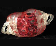
In 2002 there was a huge leap forward when CT and MRI data was used to print anatomical models of conjoined twins Mohamed and Ahmed Ibrahim. Created by Medical Modeling, the models were used for everything from creation of the custom bed to mapping of important vessels during the separation surgery completed successfully in 2003. Since then, using the technology for planning these types of very complicated surgeries is an accepted, even expected approach.
The data for the models came from CT and MRI scans. Medical Modelling President and Chief Technology Officer Andrew Christensen explains that the models were created using stereo lithography. Two sets of data were used--one to model, in a transparent polymer, the bones of the skull and another to model the interconnected blood vessels, which appear red. To illustrate the red blood vessels inside the model, Medical Modelling used a photo-initiated red dye. Each slice of the model was built twice by the laser: One pass created the clear model with a lower energy ultraviolet beam, and another pass--at a much higher UV energy level--activated the polymer with the red dye.
The twins were successfully separated on Oct. 12. Both boys are making excellent progress in their rehabilitative therapies.
Source: http://www.sme.org/memagazine/article.aspx?id=73433