Doctor: Dr. Swati Garekar (Paediatric Cardiologist)
Case: 10 Month old infant weighing 4.6kg. There is a double outlet right ventricle. Great Arteries are d-imposed; aortic valve is anterior to pulmonary valve. Pulmonary valve is hypoplastic. Branch Pulmonary artery are confluent and normal in size. There is large inlet ventricular septal defect that is separated from semilunar valve by a small chunk of conal septum.
Surgery: The patient underwent a Nikaidoh complex surgery. Two great arteries arising from the heart were disconnected & repositioned. The hole in the heart was closed.
Benefit of the 3D model: The 3D model gives the surgical team a good idea of what to expect when they open the heart ‘Nobody likes Surprises’ sometimes echocardiography doesn’t do complete justice in such complex cases.
From The Doctors Desk: In complex cases like Nikaidoh a 3d model helps to understand the routing especially in such cases where there is different intracardiac routing, we know where exactly the hole is, how the great arteries are related to each other and the hole
Case Gallery
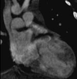 Pre Op CT Image
 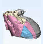 Sliced 3D printed Paediatric Heart
 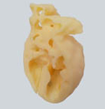 3D printed model (cross section view)
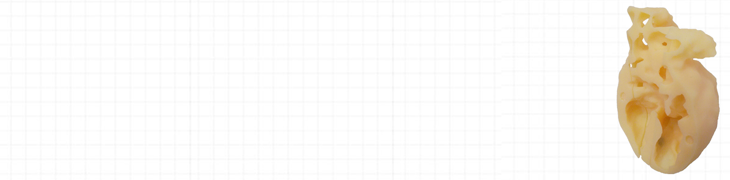
 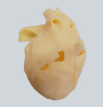 3D printed model